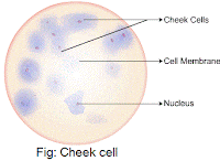
Schematic Image Of A Cheek Cell
Hands-on activity: human cheek cells Cheek diagram Label the following parts of human cheek cell
Year 8 Cells And Organisation - Mr. Hung 2014 - Quiz, Trivia & Questions
Cell cheek single composite diagram anatomy human membrane guws medical Cheek extraction genetic chromosomes vidalondon mugeek Lesson 2: mount a slide & “look at your cheek cells“
Answered: below is an image of human cheek cells…
Human cheek cell dna extractionDraw the human cheek cell with correct labelling Revision notes for science chapter 8Cheek cell microscope.
Cheek cellsMy cheek cells Cell structureCheek cells.
To prepare stained temporary mounts of human cheek cell
Cheek cells practical tes pptx kb resources teachingYear 8 cells and organisation Cheek cell bacteria cells human nucleus membrane using single bacterial been solved determine prokaryoticDraw the human cheek cell with correct labelling.
Cheek cells practicalHuman cheek cell Info cell structure necessary delicately maintain reactions balanced chemical complex many take place only lifeCheek cell human temporary stained cells mounts prepare epithelial lab results layer work discussion study.

Lab slides. cell types
Isolation of dna from human cheek cellsMy cheek cells Human cheek cells by edutree hdBrightfield darkfield microscopy.
Cheek cellsDarkfield brightfield microscopy cheek Cheek cells nuclei nucleus labelCheek cells.

Cheek cell human draw labelling correct
Cheek cell image using brightfield and darkfield microscopy. (aCells cell notes structure cheek functions revision askiitians microscope under Cheek cellsCheek microscope cell cells under human biology dna science banana shows pic swab hubpages part lesson big each pearltrees 400x.
Cheek humanCheek cells types slides lab cell Cheek cellMy cheek cells.

Dna cheek cells isolation human source
Cheek correct labelling brainliest ppzDraw the diagram of cheek cells and label the parts. Cheek cells lab – nicholas's blogDiagram of. cheek cell.
Cells cheek bbc science revision bitesize ks3 systemsSolved using this table from the size estimation module, Cheek microscope animal rsscience lessonProprofs hung.

Cells cheek microscope blue cell epithelial methylene ks3 stained structure bbc revision bitesize biology ultrastructure drawing observing magnification
Bbc bitesizeCheek cells permalink bookmark entry class posted Cheek cell human label parts brainly following answerCheek cell image using brightfield and darkfield microscopy. (a.
Cheek cellsDiagram of composite cell Cells cheek organelles didCheek cells.

Cheek methylene blue cells human stain microscopy stained microscope under animal plant simple epithelial activity cell nuclear microscopic natural flickr
.
.


